Intermittent Thoughts on Building Muscle: Estrogen, Friend or Foe of Skeletal Muscle Hypertrophy? Plus: "Hey, Bro! Are You 'SERMing' Away Your Satellite Cells?"
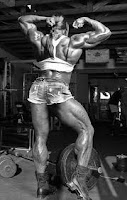 |
| Image 1: Iris Kyle's back is a living testostomy, ah... pardon testimony to the muscle building powers of estrogen ;-) |
Estrogen makes your muscles weak and your belly fat, right? Not exactly.
Completely contrary to common wisdom, estrogen is by no means the exact counterpart to testosterone. In fact, its potential facilitative if not beneficial or required effects on skeletal muscle hypertrophy are just as undebatable as the negative effects a skewed testosterone to estrogen ratio will have on both the overall health, as well as the physical appearance of men and women.
In an extensive review of the literature, Enns and Tiidus propose the following purported mechanisms by which estrogen may factor in the accrual, repair and maintenance of skeletal muscle tissue (Enns. 2010):
- estrogen as an antioxidant: previous studies have shown that low/high levels of estrogen are associated with decreases / increases in reactive oxygen specimen and markers of inflammation;
- estrogen as a membrane stabilizer: while it is probably difficult to distinguish this effect from the aforementioned antioxidant effects of estrogen, it has been shown that by intercalating within membrane
phospholipids, estrogen contributes to the stability of the cell membrane;
- downstream effects of estrogen receptor binding: dozens of studies have investigated the metabolic effects of estrogen receptor alpha and beta activation; a recent review by Barros and Gustafson (Barros. 2011), for example emphasizes its role in the well-known insulin induced expression of GLUT-4 receptors on the cell-membrane of the muscle; in that, ERα modulates GLUT4 translocation to the cell membrane and thusly stimulates glucose uptake, whereas ERβ is a repressor of GLUT4 expression; in view of what you have learned about the potentially insulin sensitizing effects of testosterone the latter could well depend on the ratio of estrogen to estrone to which the testosterone is converted in the course of central, as well as peripheral aromatization processes; with a high estradial to estrone ratio favoring insulin resistance (estradiol has identical binding affinities for both receptors, while estrone is more or less ERα specific) - a more recent studies by Rüegg et al. does yet suggest that the complete absence of ER is equally detrimental (Ruegg. 2011) and thusly corroborates assessment that our knowledge of the complex endocrine-metabolic interactions is still very limited...
Estrogen and mitochondrial biogenesis
 |
| Image 2: Does estrogen make women better endurance athletes because it increases mitochondrial biogenesis and gears your metabolism towards fatty acid not glucose oxidation? And if that is the case, would men benefit from some more estrogen, as well? |
A 2010 study from the McMasters University in Ontario, Canada (Maher. 2010), which analyzed vastus lateralis samples of 12 male and 11 female "moderatly avtive" subjects and found that
women have more protein content of the major enzymes involved in long and medium chain fatty acid oxidation which could account for the observed differences in fat oxidation during exercisewould support this hypothesis. After all, this effect could well be related to the "constantly" (de facto estrogen levels obviously vary cyclical ;-) higher amount of the skeletal muscle tissue of the female subjects is exposed to. So, the next time you are huffing and puffing on a jogging tour with your girlfriend, guys, you know that the 3 beers you had the evening before are only part of the explanation for the superior stamina of your significant other ;-)
Feminists please plug your ears: Men and women are different!
If we now remind ourselves of the initially mentioned limitations, the question arises, in how far any of the effects we have discussed so far may be sex-specific. The aforementioned example of increased fatty acid oxidation in response to estrogen mediated PGC-1a expression, for example, is supported by other researchers, like Tanopolsi (Tanopolski. 2008). Nevertheless, it does yet not bear direct experimental verification: In 2011, Salehzadeh et al. incubated myotubes (muscle fibers) from male and female donors (post-menopausal and age-matched male controls) with either testosterone or 17b-estradiol and found that male and female myotubes respond very differently to "their" respective sex hormones (Salehzadeh. 2011):
Testosterone and E(2) treatment enhanced insulin-stimulated glucose incorporation into glycogen and AKT phosphorylation in myotubes from female donors, highlighting a sex-specific role of sex hormone in glucose metabolism. Testosterone treatment increased palmitate oxidation in myotubes from both female and male donors, while E(2) enhanced palmitate oxidation in myotubes from male donors only. Testosterone-mediated increase in palmitate oxidation was attenuated at the presence of androgen receptor antagonist, which may indicate a role of nuclear steroid receptor in muscle lipid oxidation. [...] E(2) treatment increased pyruvate dehydrogenase kinase 4 mRNA expression in myotubes from female donors. Thus, our data suggest that testosterone or E(2) modulates muscle glucose and lipid metabolism and may play a role in metabolism in a sex-dependent manner.In the muscle cells of female donors, E(2) [=17b estradiol] acts similarly to testosterone, it increases protein synthesis glycogen storage and protein synthesis (the latter via the well-known AKT pathway). Contrary to what the Zorzona study would suggest, 4-day incubation with estrogen did yet fail to increase the oxidation or fatty acids and geared the energy system of the myotubes from the female donors more towards the glycolytic pathway (thus the increase in pyruvate dehydrogenase). Against that background it seems totally paradoxical that it increased the fatty acid oxidation in the myotubes from the male donors. I mean, don't we all "know" that estrogen makes you fat?
Does estrogen make you fat? Or does fatness make you estrogenic?
 |
| Image 3: Beer belly because or despite high estrogen levels? Is that the question or did we get it totally wrong? |
Another potential explanation for the lack of "anti-obesogenic" effects of estrogen could simply be its inability to "enter" the fat cells. This is an issue for almost all hormones and a way your body has developed to get water-soluble compounds into a cell is to attach a sulfur molecule to the compound. For estrogen the enzyme that catalyzes this reaction is called estrogen sulfotransferase (EST) and its exceptional high activity in male white adipose tissue has been investigated by several researchers, lately. In August 2011 Wadga et al. report that its expression in pre-adipocytes inhibits their maturation (Wagda. 2011), an observation that should remind you of the anti-adipogenic (i.e. blocking the genesis of new fat cells) of testosterone about which you have learned in the last installment. With testosterone being a "pro-hormone" to estrogen, this suggest that part of this effect could be mediated by local aromatization to estrogen. The increased adipocyte number and size Misso et al. observed in an aromatase deficient (Misso. 2003), and Ohlson et al. in a estrogen receptor alpha deficient mouse model (Ohlson. 2000) support the hypothesis that, even for men, low estrogen levels could contribute to increased body fat levels.
Estrogen inhibits the maturation of pre-adipocites, ok, but what about "pre-myocytes"?
While it may seem as if I my "intermittent train of thought" has once again lost track of the topic at hand we are actually closing in on what I belief could be one of the most important hypertrophy-specific effects of estrogen: its interaction with satellite cells. That the latter are an important factor in the myogenic equation should not be news to anyone who has been following this series over the past couple of weeks. That the sustaining effects estrogen exerts on these "pre-myocytes" could at least partly explain the drastic difference in sheer muscle mass gains, users of performance enhancing drugs notice from so called "wet" compounds.
Contrary to their "dry" counterparts, these drugs are either susceptible to the aromatase enzyme (mostly to a different degree than testosterone, though) and will consequently be partially converted to estrogen or they do exhibit a certain binding affinity for either the estrogen-alpha or -beta receptor right away (cf. "Beyond Vida" for more info on the binding affinities of various compounds).
The most relevant data (because it does not come from pre- or post-menopausal women, let alone overiectomized rodents) with regard to the beneficial effects of estrogen on skeletal muscle cells comes from a 2005 study by Tidus et al. (Tidus. 2005), who counted the number of satellite cells in a given area of myofibers of red soleus (=slow twitch, type II) and white vastus (=fast twitch, type I) muscle tissue after 90min of intermittent (5min running, 2 min rest) downhill (-13.5°) running on a rodent treadmill at 17m/min (=4.7km/h).
 |
| Figure 1: Satellite cell and neutrophil count in normal male rats and male rates who were implanted with a 25mg estrogen pallet before and 74h after 90 min of intermittent downhill running (data adapted from Tidus. 2005) |
Estrogen and satellite cell activation, proliferation and survival
The geeky smart-asses that we are, we will obviously not content ourselves with these observations. I mean, yeah... estrogen is facilitative, but is it necessary, as well? In a way it is quite ironic that it is, once again, a drug that is commonly used by steroid users which provides the answer to this question. The respective study was published in Development and Stem Cells and its title, "Effective fiber hypertrophy in satellite cell-depleted skeletal muscle" would actually suggest that it contradicts everything we have been discussing before. Therefore I deem it necessary to initially point out that the hypertophy response subsequent to synergistic ablation for 2 or 6 weeks was identical for two weeks and slightly reduced after 6 weeks, if we only consider the muscle weight. If, however, we take a closer look at the myofibrial structure, we see the same, in the longer term unsustainable or pathological increases in domain sizes we have discussed in many of the previous installments (e.g. "Growing Beyond Physiological Limits").
 |
| Figure 2: Number of myofibers of different sizes in control and mice exposed to synergistic ablation surgery (gastrocnemius and soleus) after two weeks (left) and percent of myofibers with central nuclei (right; data adapted from McCarthy. 2011) |
Hey bro! Are you SERMing away your growth potential?
That estrogen, or I should say the proper activation of the estrogen receptors, is necessary for the maintenance of adequate satellite cell levels, even in the absence of exercise induced muscle damage and consequent satellite cell recruitment, becomes evident, when we take a closer look at the way the scientists depleted the satellite cell pool of their mice (note: while these were female mice, Lepper et al. used the same method in male mice, cf. Lepper. 2011): They used tamoxifen!
 |
| Figure 3: Satellite cell count in muscles of mice after treatment with vehicle or 2mg/day of tamoxifen for five consecutive days (left) and images of stained and marked gastrocnemius samples (data and images adapted from McCarthy. 2011) |
Assuming that most of you will be aware that tamoxifen (brand name Nolvadex), the hepatoxic effects of which I have addressed in a recent blogpost, is a selective estrogen receptor modulator (SERM), or in other words a synthetic molecule that binds to the estrogen receptor without activating it, it should be obvious that without estrogen, or any other substance that would "dock" to and activate the estrogen receptor healthy, continuous and sustainable muscle growth is impossible.
Take home message: Estrogen is necessary for continuous and sustainable muscle growth
Now, while the take home message that estrogen is in fact a necessary prerequisite of skeletal muscle hypertrophy (at least in the long run), this observation brings the previously raised question in how far the "muscle building effects" of exogenous testosterone, as they were for example observed in healthy young men by Bhasin et al. (Bhasin. 2001) and in community dwelling elderly men on testosterone-replacement therapy by Shinha-Hikim et al. (Shinha-Hikim. 2006) are not, on a cellular level, at least partly mediated by the aromatization of testosterone to estrogen.
And as if things were not already complicated enough, testosterone is a "pro-hormone" not only to estrogen, but also to dihydrotestosterone (DHT), of which bro-science would have it that it is a 10x more potent androgen than the "Big T", itself. As you are probably suspecting by now, we will have to postpone the discussion of the involvement of the manliest of all androgens to the next installment of the Intermittent Thoughts ;-)


